2D DNA-origami assembly for Cryo-EM Applications


We propose a self-assembled DNA origami honeycomb 2D-lattice as a molecular imaging scaffold for Cryo-Electron Microscopy (Cryo-EM). The thin, micrometer-scale supporting scaffold is able to cover extended areas of the holey carbon film and sufficiently resilient to withstand blotting and plunge-freezing forces. Furthermore, the DNA binding sites can be chemically engineered for selective surface-affinity trapping. We demonstrate the advantages of the method to facilitate membrane vesicles sample preparation. This includes increasing the local density of the vesicles, enabling the study of low-abundance membrane complexes, and purifying heterogenous samples by isolating vesicles from cell conditioned medium.

Free-Standing DNA Origami Superlattice to Facilitate Cryo-EM Visualization of Membrane Vesicles
Nesrine Aissaoui*, Allan Mills*, Josephine Lai-Kee-Him*, Nicolas Triomphe, Quentin Cece, Christine Doucet, Anne Bonhoure, Michel Vidal, Yonggang Ke, and Gaetan Bellot.
 

GaetanJACS 1000px

When cryo-electron tomography uncovers the in cellulo structural organization of the Chikungunya virus replication complex


When cryo-electron tomography and sub-tomogram averaging techniques uncover the in cellulo structural organization of the Chikungunya virus replication complex within the plasma membrane


Bron chikv 16x10


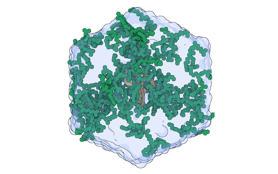
The Chikungunya virus (CHIKV) is a mosquito-borne pathogen responsible for acute musculoskeletal disease in humans. The replication of the viral RNA genome occurs within specialized membranous replication organelles (RO), also known as spherules, which house the viral replication complex. This membranous complex consists of four viral proteins (nsP1-4) along with various cellular partners.

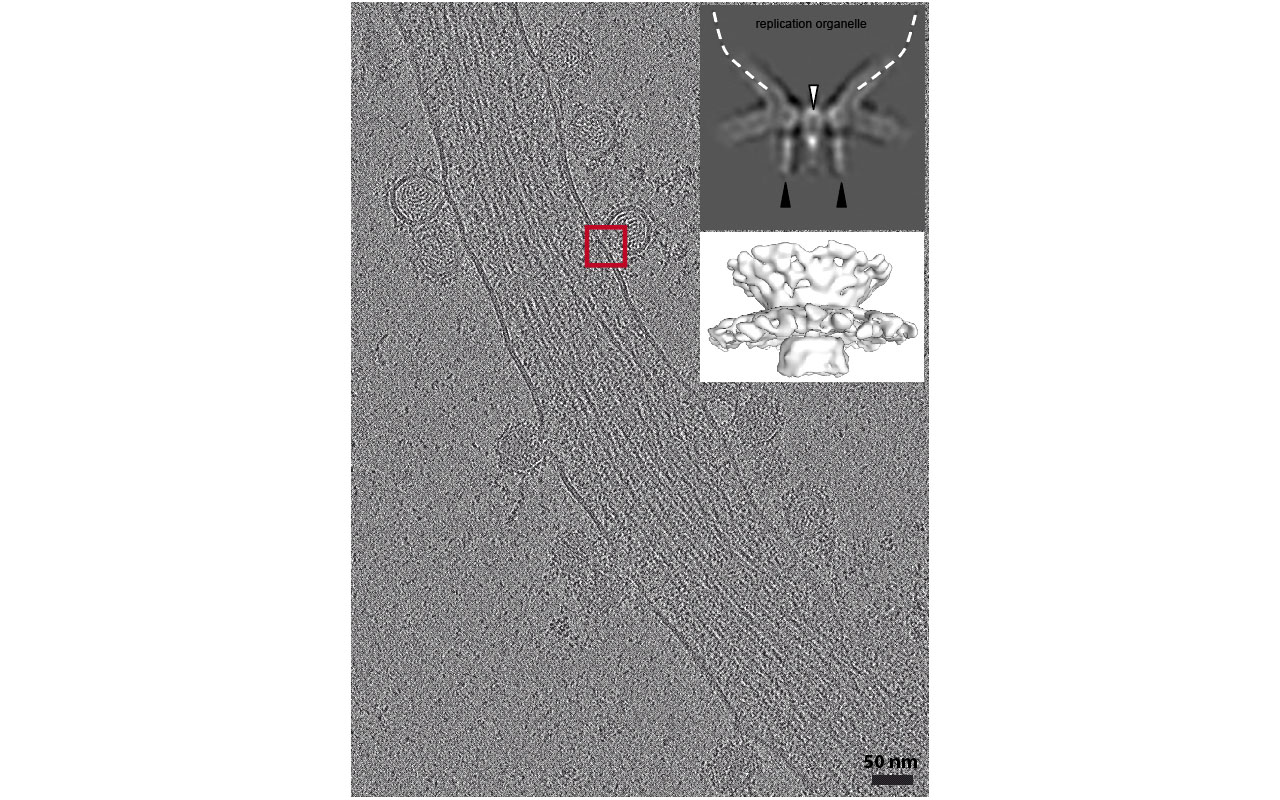
Using a combined approach of cryo-electron tomography on infected human cells at 17 hours post-infection (hpi), sub-tomogram averaging, and cryo-sectioning through CEMOVIS (Cryo-Electron Microscopy of Vitreous Sections), we investigate the structural organization of the replication complex and examine the replication dynamics within the spherules.

Collectively, this study sheds new light on the dynamic behavior of CHIKV replication organelles and the associated viral replication processes at the interface with cell membranes in infected cells.


Publication link

In situ fate of Chikungunya virus replication organelles

Justine Girard, Olivier Le Bihan, Joséphine Lai-Kee-Him, Maria Girleanu, Eric Bernard, Cedric Castellarin, Matthew Chee, Aymeric Neyret, Danièle Spehner, Xavier Holy, Anne-Laure Favier, Laurence Briant and Patrick Bron

J Virol. 2024 Jun 28:e0036824.doi: 10.1128/jvi.00368-24

New Structure of a Protein Modulating Bacterial Gene Silencing

Antibiotic resistance and the appearance of new virulent bacterial strains constitute a major threat to human health. The problem is aggravated by the transfer of resistance and virulence genes between bacteria (horizontal gene transfer). In this context, a detailed knowledge of the mechanisms allowing bacteria to tolerate the acquisition of foreign DNA is lacking and it may open the way to new sustainable strategies to fight infectious diseases infectious diseases.

Here we describe a structural model for the complex between Hha and H-NS proteins which selective represses genes in Enterobacteria acquired by horizontal transfer. We found a charge zipper formed by interdigitation of residues from three proteins stabilizes the complex. Charge zippers provide selectivity to electrostatic protein complexes and understanding selective gene silencing may help fighting antibiotic resistance

New publication: "A Three-protein Charge Zipper Stabilizes a Complex Modulating Bacterial Gene Silencing"
Authors: Tiago N. Cordeiro, Jesús García, Pau Bernadó, Oscar Millet et Miquel Pons
Journal: Biol Chem. 2015 Aug 28;290(35):21200-12
Link to this article

Insights into the "cocktail effect" of endocrine disruptors

Chemicals which taken in isolation are safe for humans may become harmful when mixed . The team of William Bourguet in Structural Biochemistry Center (Inserm / CNRS / University of Montpellier), together with teams from the Cancer Research Institute (IRCM ) and the Functional Genomics Institute (IGF ) in Montpellier elucidated in vitro a molecular mechanism that may contribute to this phenomenon known as the "cocktail effect".

New publication: "Synergistic activation of human pregnane X receptor by binary cocktails of pharmaceutical and environmental compounds"
Authors: Delfosse V, Dendele B, Huet T, Grimaldi M, Boulahtouf A, Gerbal-Chaloin S, Beucher B, Roecklin D, Muller C, Rahmani R, Cavaillès V, Daujat-Chavanieu M, Vivat V, Pascussi JM, Balaguer P, Bourguet W.
Journal: Nat Communication 2015 Sep 3;6:8089.

Links:

Activation Mechanism of the Morphine Receptor Revealed

A close collaboration between the team of Sebastien Granier (IGF) in Montpellier, Hélène Déméné (CBS, team "NMR, Structure, Dynamics and Function of Biomolecules by NMR") and the team of Prof. Brian Kobilka of Stanford University, Nobel Prize in Chemistry in 2012, helped to decipher the molecular mechanisms of activation of the morphine receptor. This receptor, also called mu opioid receptor (μOR) was investigated using combining X-ray crystallography and Nuclear Magnetic Resonance (NMR). The morphine receptor is a membrane protein belonging to the family of G protein coupled receptors (GPCR) whose operating mechanism is not well known. This lack of knowledge is partly due to the difficulties encountered in the investigation of these membrane proteins by structural biology approaches. This study has been published in the scientific journal Nature (Sounier et al, Nature, 524 (7565). -doi 375-8: 10.1038 / nature14680).

The NMR study reveals how the binding of a ligand that mimics morphine (agonist) induces changes in conformational states of μOR from an inactive state to an active state. This active state is the only one capable to lead to intracellular signal transduction via activation of signaling proteins such as G protein. The study also shows that the active state can be achieved only if the agonist and G protein are linked to the receiver simultaneously. This property was previously observed for only one of the 800 members that make up the family of GPCRs. Thus, the study reveals how the activation signal propagates through the different areas of the receptor and proposes that this process plays a key role in signal transduction. Overall, these data shed light on the little known process of activation of GPCRs and its dynamics.

New publication: "Propagation of conformational changes during μ-opioid receptor activation" 
Authors: Sounier R, Mas C, Steyaert J, Laeremans T, Manglik A, Huang W, Kobilka BK, Déméné H, Granier S.
Journal: Nature. 2015 Aug 20;524(7565):375-8. doi: 10.1038/nature14680.

Link to this article

Connexion