Engineering ‘Plug-and-Play’ Cell-Free Biosensors

Cell-free protein synthesis represents a promising avenue for developing next-generation diagnostics. Researchers at the Center for Structural Biology in Montpellier and the Michalis Institute in Jouy-en-Josas recently expanded the potential of these 'cell-free biosensors' by combining transcription factor-based detection with metabolic engineering. The synthetic biosensors are capable of detecting benzoic acid in commercial beverages and hippuric acid and cocaine in human urine. These results have been published in the journal Nature Communications along with a 'Behind the Scenes' article for the Nature Bioengineering Community.

cell free biosensors

Transcription and chromosome observation in single cells with Hi-M

Direct and simultaneous observation of transcription and chromosome architecture in single cells with Hi-M

Andrés M. Cardozo Gizzi, Sergio M. Espinola, Julian Gurgo, Christophe Houbron, Jean-Bernard Fiche, Diego I. Cattoni, Marcelo Nollmann

Simultaneous observation of 3D chromatin organization and transcription at the single cell level and with high spatial resolution may hold the key to unveil the mechanisms regulating embryonic development, cell differentiation and even disease. We have recently developed Hi-M, a technology that allows for the sequential labelling, 3D imaging and localization of multiple genomic DNA loci together with RNA expression in single cells within whole, intact Drosophila embryos. Importantly, Hi-M enables simultaneous detection of RNA expression and chromosome organization without requiring sample unmounting and primary probe re-hybridization. Here, we provide a step-by-step protocol describing the design of probes, the preparation of samples, the stable immobilization of embryos into microfluidics chambers, and the complete procedure for image acquisition. The combined RNA/DNA fluorescence in situ hybridization procedure takes 4-5 days including embryo collection. In addition, we describe image analysis software to segment nuclei, detect genomic spots, correct for drift and produce Hi-M matrices. A typical Hi-M experiment takes 1-2 days to complete all rounds of labelling and imaging and 4 additional days for image analysis. This technology can be easily expanded to investigate cell differentiation in cultured cells, or organization of chromatin within complex tissues.

Nat Protoc 2020 Mar;15(3):840-876. doi: 10.1038/s41596-019-0269-9

NatProt2020

ATP-driven separation of liquid phase condensates in bacteria

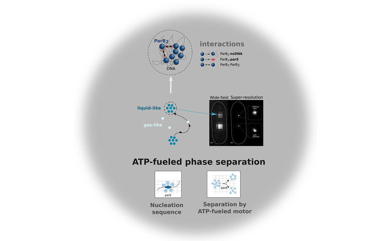
Liquid-liquid phase separated (LLPS) states are key to compartmentalise components in the absence of membranes, however it is unclear whether LLPS condensates are actively and specifically organized in the sub-cellular space and by which mechanisms. Here, we address this question by focusing on the ParABS DNA segregation system, composed of a centromeric-like sequence (parS), a DNA-binding protein (ParB) and a motor (ParA). We show that parS-ParB associate to form nanometer-sized, round condensates. ParB molecules diffuse rapidly within the nucleoid volume, but display confined motions when trapped inside ParB condensates. Single ParB molecules are able to rapidly diffuse between different condensates, and nucleation is strongly favoured by parS. Notably, the ParA motor is required to prevent the fusion of ParB condensates. These results describe a novel active mechanism that splits, segregates and localises non-canonical LLPS condensates in the sub-cellular space.

B. Guilhas, J.C. Walter, J. Rech, G. David, N.-O. Walliser, J. Palmeri, C., Mathieu-Demaziere, A. Parmeggiani, J.Y. Bouet, A. Le Gall1, M. Nollmann

Full text: https://doi.org/10.1101/791368

CNRS news: https://insb.cnrs.fr/fr/cnrsinfo/comment-la-separation-de-phases-aide-la-segregation-du-materiel-genetique


MolCell2020

Study on cis-regulatory chromatin loops

Cis-regulatory chromatin loops arise before TADs and gene activation, and are independent of cell fate during development

Sergio Martin Espinola, Markus Götz, Jean-Bernard Fiche, Maelle Bellec, Christophe Houbron, Andrés M. Cardozo Gizzi, Mounia Lagha, Marcelo Nollmann

During development, naïve cells gradually acquire distinct cell fates, through sophisticated mechanisms of precise spatio-temporal gene regulation. Acquisition of cell fate is thought to rely on the specific interaction of remote *cis*-regulatory modules (e.g. enhancers, silencers) (CRM) and target promoters. However, the precise interplay between chromatin structure and gene expression is still unclear, particularly in single cells within multicellular developing organisms. Here we employ Hi-M, a single-cell spatial genomics approach, to systematically detect CRM-promoter looping interactions within topological associating domains (TADs) during *Drosophila* development. By comparing *cis*-regulatory loops in alternate cell types, we show that physical proximity does not necessarily instruct transcriptional states. Moreover, multi-way analyses revealed the existence of local interactions between multiple remote CRMs to form hubs. We found that loops and CRM hubs are established early during development, prior to the emergence of TADs. Moreover, CRM hubs are formed via the action of the pioneer transcription factor Zelda and precede transcriptional activation. Our approach offers a new perspective on the role of CRM-promoter interactions in defining transcriptional activation and repression states, as well as distinct cell types.

https://www.nature.com/articles/s41588-021-00816-z

Cis-reg-loops

Important implication of intrinsically disordered protein in signaling pathways

Cellular signaling machinery is involved in major physiological processes and therefore in a large number of diseases. However, full understanding of these complex systems function and deregulation, in a physiopathological context, is made difficult by the presence of a large number of disordered regions. Their structural and functional properties are still largely unexplored. In this chapter, we present the signaling pathways of G protein-coupled receptors and those of nuclear receptors. We describe the involvement of these disordered regions of the different actors which are at the heart of the action mechanism.


Myriam Guillien, Albane le Maire, Assia Mouhand, William Bourguet, Jean-LouisBanères, Nathalie Sibille*


Full Text: https://doi.org/10.1016/bs.pmbts.2020.05.001


arrestin gpcr

Connexion