Plateau de Biophysique (Biophys)
Responsable scientifique à contacter : Cette adresse e-mail est protégée contre les robots spammeurs. Vous devez activer le JavaScript pour la visualiser.
Responsables techniques : Cette adresse e-mail est protégée contre les robots spammeurs. Vous devez activer le JavaScript pour la visualiser. , Jean-Bernard Fiche
Matériels techniques
Etude type en Biophysique
o 1 : Objectif
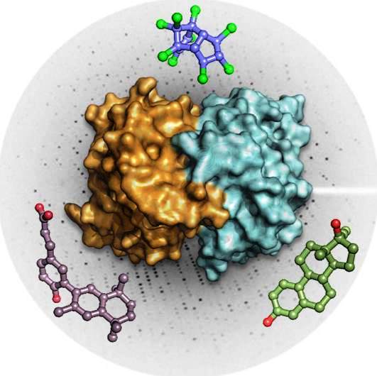
Cette plateforme propose un ensemble d'approches basées sur la spectroscopie de fluorescence applicables à des études d'interactions entre biomolécules et de dynamique structurale (repliement, assemblage, diffusion, fluctuations, changements conformationnels). Contactez Emmanuel Margeat ou Caroline Clerte pour discuter des possibilités d'utilisation de la plateforme et de la conception des expériences.
Ci dessous une présentation des différentes techniques utilisées au CBS (bientôt disponible).
Plateau de microscopie en champ proche (AFM)
Scientific manager : Cette adresse e-mail est protégée contre les robots spammeurs. Vous devez activer le JavaScript pour la visualiser.
Technical manager: Cette adresse e-mail est protégée contre les robots spammeurs. Vous devez activer le JavaScript pour la visualiser.
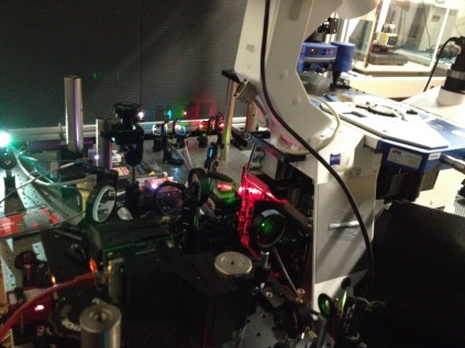
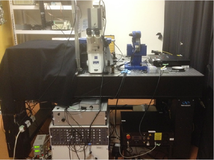
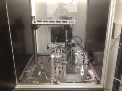
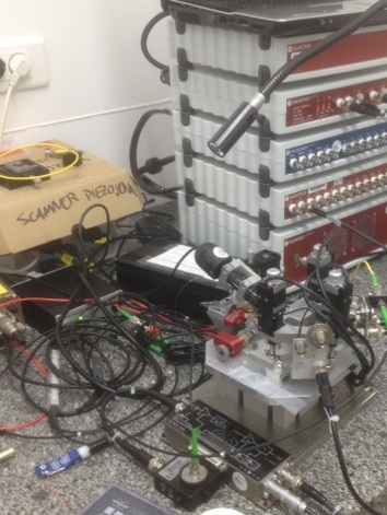
Instruments and techniques:
 |
 |
 |
|
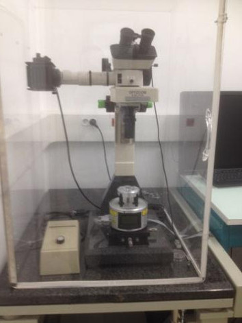
JPK Nanowizard 4 coupled to Super Resolution Fluorescence Microscopy
|
JPK Nanowizard coupled to
Confocal Microscopy, PIE-FRET and FCS
|
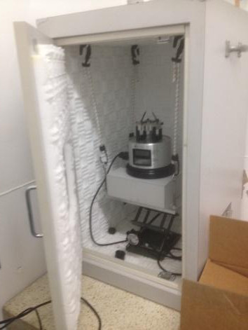
High-Speed Atomic Force Microscope
(Developed in collaboration with Pr.Toshio Ando)
|
 |
 |
 |
|
Bruker Multimode Nanoscope VIII
|
Bruker multimode Nanoscope III
|
X-Ray-AFM that can be installed in Synchrotron X-Ray beamlines. Equiped with complete Nanonis SPECS control unit.
|
Atomic Force Microscopy (AFM):
The technique allows characterizing sample morphology at nanoscale. Images are acquired by scanning a nanometric tip in gentle contact or intermittent contact with the sample. The tip is positioned at the end of a micrometric force transducer, the AFM cantilever: it records the variation of sample topography due to changes of tip-sample interaction during scanning operation. In addition, by measuring the tip-sample interaction force as a function of the tip-sample distance, AFMs can evaluate sample elastic and viscous properties.
Objectives:
- Characterize biological samples topography at nanoscale in dry or liquid environment. Image acquisition can be performed in static or dynamic modes.
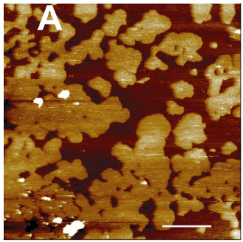
Cholera Toxin B-oligomers (left) bound to GM1 domains within a DOPC-DPPC (1:1) model membrane (right) as observed by AFM. Milhiet, Pierre Emmanuel, et al. "AFM characterization of model rafts in supported bilayers." Single molecules 2.2 (2001): 109-112.
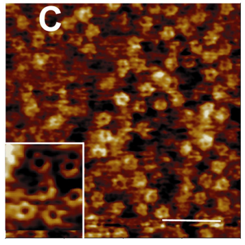
- Correlate biomolecules activity observed by Fluorescence (Epi and TIRF) or Super Resolution Microscopy (PALM/STORM) with the associated sample topology observed by AFM. Our two fluorescence-AFM correlative setups are mounted on top of Zeiss inverted microscopes. Fast AFM imaging (1 frame in few seconds) can be performed simultaneously with the fluorescence microscopy operation.
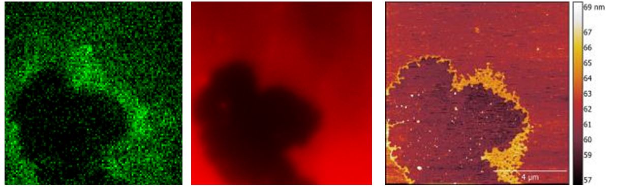
Amphiphatic Lipid Packing Sensors (ALPS, left), and DOPE (Rhodamin marked, center) TIRF image correlated with the AFM image of the model membrane + biomolecules on the right. Scan size = 10 µm.
- Real-time visualization of biomolecules activity at high spatial-temporal resolution by High-Speed AFM (HS-AFM). Our HS-AFM can acquire AFM images up to 10 images/seconds, allowing for dynamic tracking of slow biomolecules activity.
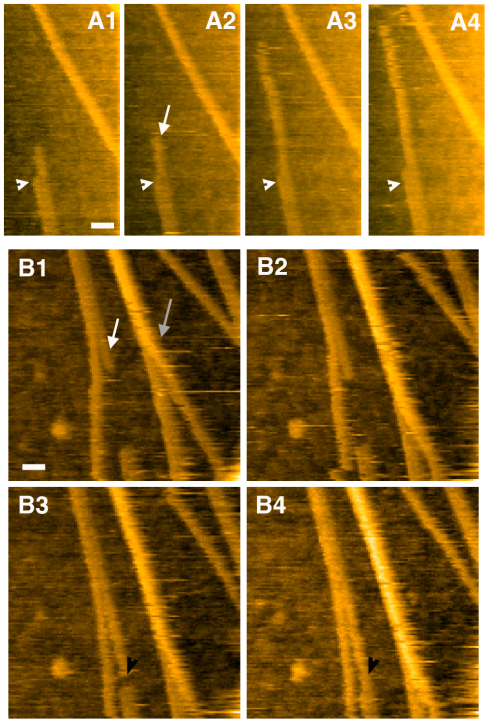
Elongation of single protofibril (white arrows) is shown in time lapses. Deciphering the Structure, Growth and Assembly of Amyloid-Like Fibrils Using High-Speed Atomic Force Microscopy. Milhiet, P. E., Yamamoto, D., Berthoumieu, O., Dosset, P., Le Grimellec, C., Verdier, J. M., ... & Ando, T. (2010). PLoS One, 5(10), e13240. Speed = 1 image/second.
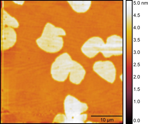
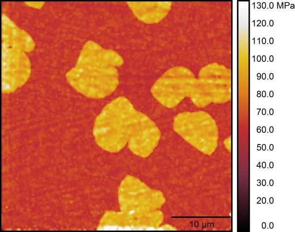
- Evaluation of mechanical properties of biomolecules and cells. Making use of the AFM tip as a nanometric indenter, we can characterize the extrinsic and, if possible, the intrinsic rigidity of materials with high spatial resolution.
Left: DOPC-DPPC (1:1) model membrane morphology. Right: corresponding Young modulus evaluated modeling the tip-sample mechanical contact with the Hertz theory.
Software and analysis:
We make use of both commercial and entirely/partial custom-made software to control our instruments. Data analysis is performed both with C++/Matlab home-made programs as well as with open source software such as ImageJ and Gwyddion or software provided by manufacturers.
")
")


![Rejoignez le programme de master qbio [Quantitative Biology : Mécanismes moléculaires des systèmes vivants (parcours IDIL)] Session de questions-réponses le lundi 10 février !](https://www.cbs.cnrs.fr/cache/mod_bt_contentslider/3e10f899c82679e1992f2a701c367d27-qbio_IDIL_quadri_16x10.png "Rejoignez le programme de master qbio [Quantitative Biology : Mécanismes moléculaires des systèmes vivants (parcours IDIL)] Session de questions-réponses le lundi 10 février !")
 de Montpellier s’engage à accueillir 6 stagiaires de seconde générale et technologique, du 16 au 27 juin 2025")