Memberships
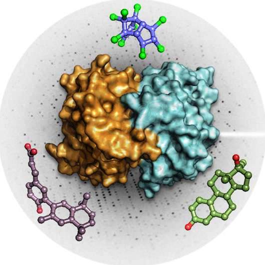
The French Infrastructure for Integrated Structural Biology (FRISBI) provides an infrastructure for integrative structural biology approaches, from the molecular to the cellular level, integrating multi-resolution data from X-ray crystallography, small angle X-ray scattering, NMR, Cryo-EM and functional data including development for protein expression and crystallization. FRISBI is open to structural and molecular and cell biologists from both academia and industry from France and Europe.

France‐BioImaging is a large‐scale national research infrastructure. France-Bioimaging will deploy a distributed biological imaging infrastructure in a coordinated and harmonized manner. At the frontier between molecular and cell biology, biophysics and engineering, mathematics and bioinformatics, France-BioImaging gathers, several outstanding biological Imaging Centerssupported by state of the art R&D teams with the aim to cover recent advances in microscopy, spectroscopy, probe engineering and signal processing. Thereby France-BioImaging will provide quantitative measurements, computational analysis and an integrative understanding of a wide range of cellular and tissular activities.
France-BioImaging is a highly pluridisciplinary project with participants in Biology, Physics, Chemistry, Computer science and engineering. The strength of the France-BioImaging consortium is to put efforts together to overcome technological barriers persisting at different levels of Cellular Imaging. Different solutions for each challenge are proposed among different nodes, justifying a second level of France BioImaging organization as shared technological and methodological Work Packages.
More information on the France BioImaging website

By ministerial decision of May 8, 2018, ChemBioFrance is created and registered on the national roadmap of research infrastructures. ChemBioFrance gathers four different ressources and networks: the national chemical library, a network of screening platforms, a distributed chemoinformatics platform, a network of ADME platforms.
Its management is entrusted to the Institute of Chemistry (INC) of the Centre National de la Recherche Scientifique (CNRS), which delegates management to a Research Federation: ChemBioFrance.
It offers « smart »chemical libraries with high potential for bioactivity, partnership with chemists for medicinal chemistry, drug screening services, tools for data analysis and data mining in chemical collections, ADME and toxicology services for the characterization and development of new biologically active molecules.
Its activities include :
- Conception and analyzing of chemical library for screening.
- Analyzing of screening gross data.
- Virtual screening (similarity 2D and 3D, pharmaceutical library, docking).
- Biological profiling of molecules (target prediction).
- Calcul/ Prediction of physicochemical properties (e.g: solubility, logP)
More information on the ChemBioFrance website
BioCampus Montpellier is the service unit of the RABELAIS BioHealth Cluster (Pôle Biologie-Santé). This unit is jointly funded and managed by CNRS (UMS 3426), INSERM (US 09), and Montpellier University. BioCampus Montpellier manages the main technological platforms dedicated to Life Science research in the Mediterranean part of the French Occitanie region. Beside the BioHealth domain, BioCampus Montpellier is open to the entire scientific community, particularly to biotechnology companies, pharmaceutical research, agricultural sciences and ecology, and environmental sciences.
The GIS (Scientific Interest Group) IBiSA (Infrastrutures en Biologie Sante et Agronomie) was created in May 2007. The current members of the GIS are INSERM, CNRS, INRA, CEA, INRIA, 'INCa (National Cancer Institute), the CPU (Conference of University Presidents), and the two directorates of the Ministry of Higher Education and Research, the DGRI and the DGES. The Presidency of the GIS is ensured by Yves Lévy.
It coordinates the national labeling policy and support for Platforms and Infrastructures in Life Sciences. With this in mind, GIS is taking up the policy of labeling and supporting technical personnel for the Platforms previously carried out by the Inter-Organism Meeting (RIO), and is piloting calls for projects intended to equip these platforms or to enable them to promote implementation of new technologies.
It promotes the establishment of structures for consultation and management of platforms at the regional level, which are intended to become privileged interlocutors of GIS IBiSA in the field.
It promotes animation activities (schools, thematic workshops, etc.) around the activity of the platforms.
For each of these missions, GIS launches calls for tenders.


