| Service |
AFM imaging at high resolution in liquid
|
|
| Goal |
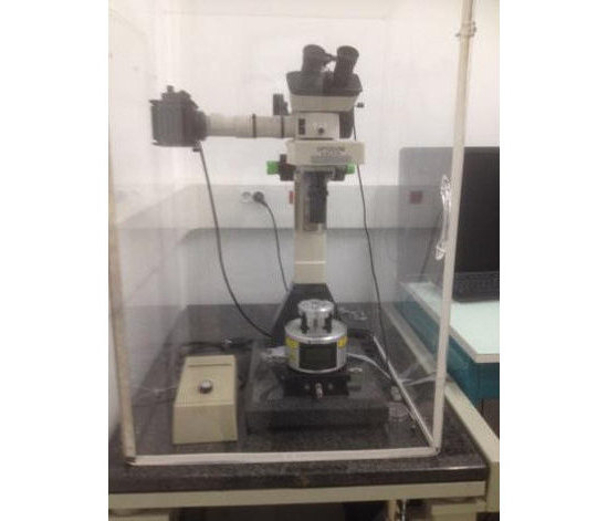
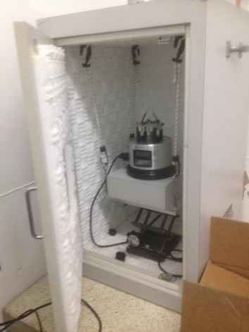
Characterize biological samples topography at nanoscale in dry or liquid environment. Image acquisition can be performed in static or dynamic modes |
|
|
The technique allows characterizing sample morphology at nanoscale. Images are acquired by scanning a nanometric tip in gentle contact or intermittent contact with the sample. The tip is positioned at the end of a micrometric force transducer, the AFM cantilever: it records the variation of sample topography due to changes of tip-sample interaction during scanning operation. In addition, by measuring the tip-sample interaction force as a function of the tip-sample distance, AFMs can evaluate sample elastic and viscous properties. |
||
|
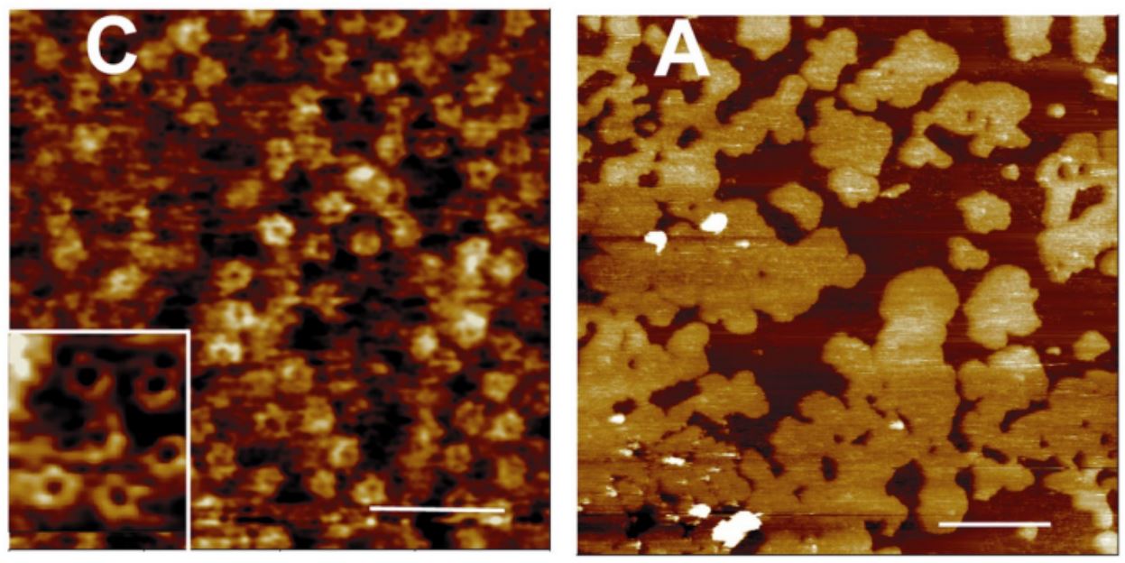
Cholera Toxin B-oligomers (left) bound to GM1 domains within a DOPC-DPPC (1:1) model membrane (right) as observed by AFM. Milhiet, Pierre Emmanuel, et al. "AFM characterization of model rafts in supported bilayers." Single molecules 2.2 (2001): 109-112 |
||
| Equipement |
