| Service |
(Cryo) Electron tomography
|
|
| Goal |
Structural analysis from molecular to a cellular level |
|
|
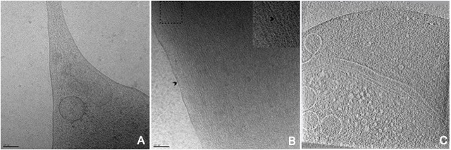
Electron tomography is a non-invasive, high resolution imaging technique that allows the visualisation of 3D organization of eukaryotic cells, with their dynamic organelles, cytoskeletal structures and molecular machines when combined with a method of arresting cells in their momentary state of function. Cellular structural biology is now becoming accessible by combining together different structural techniques. In this context, one of our goals is to investigate structure, dynamics, transmission and selectivity of viral complexes from molecular to cellular levels. |
||
|
|
||
| Comment | Need to prepare cells on EM grids | |
| Equipement |
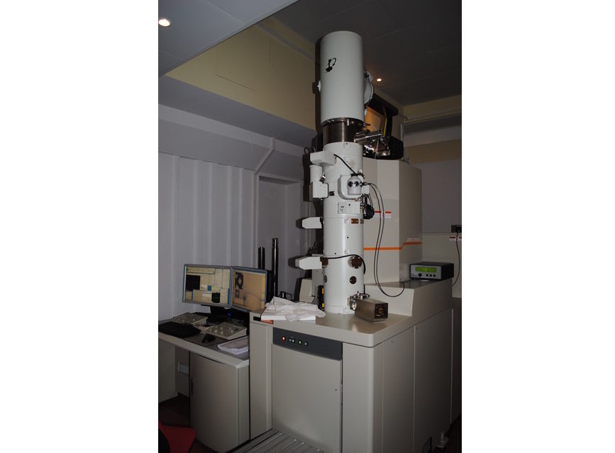
Cryo Electron Microscope
|
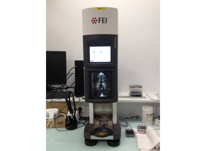
Vitrobot (ThermoFischer)
|
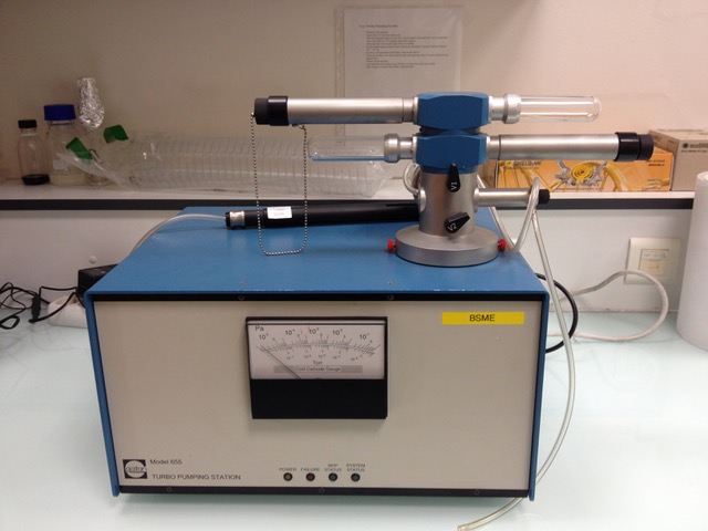
Pumping Station (Gatan) for cryo holders
|
||||||
 |
 |
 |