Micro electron diffraction

Service

Micro electron diffraction

Goal

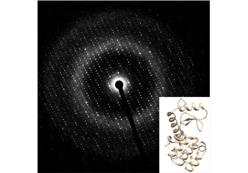
Structural analysis of tiny crystals by electron diffraction

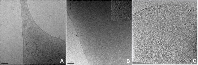
Micro electron diffraction is a recently developed method in cryo electron microscopy that allow the collection of high resolution electron diffraction data from extremely small three-dimensional crystals that are in the range of 0,1 -0,4 mm thick using a transmission electron microscope. We implemented this approach on the JEOL 2200FS electron microscope and investigated its application to membrane protein crystals.


Electron diffraction pattern of Lysozyme micro-crystal and its computed structure

 
Comment Need to prepare crystals (see X-ray crystals preparation) then cryo EM sample freezing.
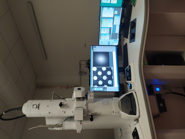
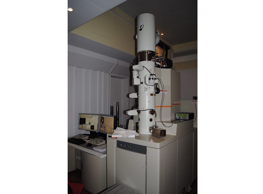
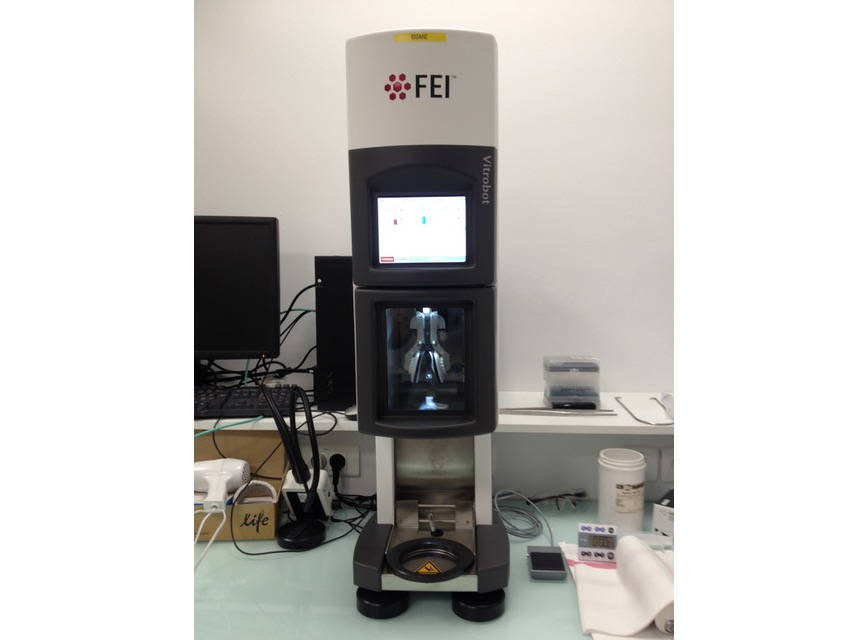
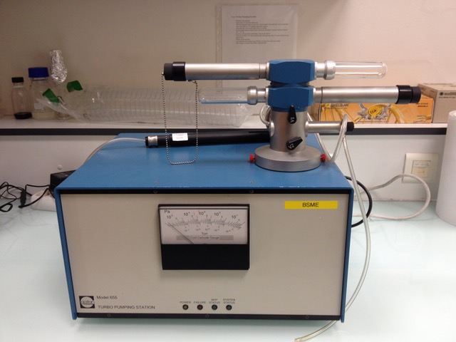
Equipement

Cryo Electron Microscope


Vitrobot (ThermoFischer)


Pumping Station (Gatan) for cryo holders


Read more ...
 
Read more ...
 
Read more ...
       

Cryo Electron Tomography

Service

(Cryo) Electron tomography

Goal

Structural analysis from molecular to a cellular level

Electron tomography is a non-invasive, high resolution imaging technique that allows the visualisation of 3D organization of eukaryotic cells, with their dynamic organelles, cytoskeletal structures and molecular machines when combined with a method of arresting cells in their momentary state of function. Cellular structural biology is now becoming accessible by combining together different structural techniques. In this context, one of our goals is to investigate structure, dynamics, transmission and selectivity of viral complexes from molecular to cellular levels.


A and B : micrographs of frozen jurkat cells, where you can see the membrane, vesicles and the actin filaments. C : CryoEM-tomogram section of HeLa cells.

 
Comment Need to prepare cells on EM grids
Equipement

Cryo Electron Microscope


Vitrobot (ThermoFischer)


Pumping Station (Gatan) for cryo holders


Read more ...
 
Read more ...
 
Read more ...
       

Analysis or screening of negative stained sample

Service

Analysis or screening of negative stained sample

Goal

Routine check of your samples

After purification of the protein complexe of interest, a negative stained grid of the sample is done. You can use this microscope to check your sample quality. It is a very easy and fast way to tell if the protein solution is homogenous, stable and a suitable candidate for high resolution structure analysis in cryo. You can also use this microscope to do room temperature tomography analysis.


Negative stainMicrograph of V2 protein complexes and 2D classes, DNA origami structures and 2D classes and cells section.

 
Comment Use this microscope as a routine instrument for the first step of you sample analysis.
Equipement

JEOL 1400 Flash Microscope

               
Microscope JEOL 1400
Read more ...
               

Single Particles Analysis

Service

Structural analysis of frozen hydrated single particles

Goal

Structure of protein complexes at high resolution

After purification of the protein complexe of interest, a negative stained grid of the sample is done. It is a very easy and fast way to tell if the protein solution is homogenous, stable and a suitable candidate for high resolution structure analysis. Then, the best protocol is used to prepare the frozen hydrated samples (good spreading of the protein complexe on the cryo grids). Images are then acquired (more than thousands images). A selection of the best images is done and 2D and 3D analysis are performed on each protein complexe particles (usually close to millions of particles).


Micrograph of ribosomes particles and the corresponding 3D structure

 
Comment Have a high quality purified sample of the protein complexe of interest (buffer composition)
Equipement

Cryo Electron Microscope


Vitrobot (ThermoFischer)


Pumping Station (Gatan) for cryo holders


Read more ...
 
Read more ...
 
Read more ...
       

Connexion