Tournants et bifurcations, acteurs de la vaso­occlusion drépanocytaire?

La drépanocytose affecte plus de 50 millions de personnes dans le monde et se caractérise par des crises douloureuses de vaso­occlusion dont la localisation exacte dans la microcirculation des tissus profonds reste inconnue. Une étude microfluidique et numérique réalisée par trois équipes multidisciplinaires de Montpellier, démontre pour la première fois que des cellules sanguines se déposent préférentiellement aux tournants à fortes courbures, et notamment aux bifurcations, caractéristiques de la microcirculation. Ces travaux qui mettent en évidence le rôle majeur que pourrait jouer la géométrie de l’écoulement dans l’avènement des crises occlusives, sont publiés dans la revue Biophysical Journal.


La drépanocytose est une hémoglobinopathie héréditaire qui affecte plus de 50 millions de personnes dans le monde. Elle se caractérise par des crises douloureuses vaso­occlusives dans la microcirculation des tissus profonds. Les symptômes proviennent d'une mutation singulière produisant une hémoglobine anormale nommée HbS dans les globules rouges. Lorsqu'elles sont désoxygénées, ces molécules d'HbS polymérisent en fibrilles traversant le globule rouge de part en part, en le déformant au repos sous une forme tristement célèbre de faucille, donnant son nom à la maladie. La perte de déformabilité résultante serait responsable d'évènements vaso­occlusifs récurrents et délétères. Au cours des trois dernières décennies, la physiopathologie des crises vaso­occlusives s'est avérée plus complexe que l'explication logique, mais trop simpliste, d'un colmatage des vaisseaux sanguins par encombrement de globules rouges rigidifiés après relargage de leur cargo d'oxygène et polymérisation de leur hémoglobine. Notamment, des scénarios complémentaires basés sur des variations permanentes des propriétés de la membrane de ces globules, telle que leur adhérence accrue entre eux et aux parois vasculaires, ont été proposés. Des cascades adhésives de globules rouges pourraient produire de la vaso­occlusion par accumulation catastrophique dans les veinules post­capillaires, même si aucune description détaillée de la dynamique d'occlusion n'est encore disponible à ce jour. La déposition et l'agrégation des globules rouges seraient en fait favorisées par un état inflammatoire chronique chez les patients souffrant de la drépanocytose, état résultant du stress oxydatif associé à l'hémolyse plus fréquente de leurs globules rouges plus fragiles. Jusqu'à présent, aucun des scénarios proposés n'a examiné le rôle éventuel joué par la géométrie de l'écoulement sur la déposition et la question de l'emplacement exact des événements occlusifs à l'échelle de la microcirculation reste ouverte. Des chercheurs du Laboratoire Charles Coulomb, de l’Institut de Mathématiques, de l’Institut de Biochimie Structurale et de Modélisation et du Laboratoire d’Hématologie de la Faculté de médecine de Montpellier, révèlent pour la première fois, grâce à l’utilisation de puces microfluidiques, que des agrégats cellulaires se forment par déposition successive de globules rouges drépanocytaires, préférentiellement autour de coins prononcés de piliers triangulaires ou de bifurcations. Cette accumulation localisée conduit à des agrégats géants flottant au milieu du canal. Puisque la microcirculation est formée de vaisseaux présentant généralement de fortes courbures, des formes ondulées, et un grand taux de bifurcations avec des longueurs de segments courts entre les bifurcations du réseau, les chercheurs proposent que ces caractéristiques géométriques pourraient jouer un rôle majeur dans le développement d'évènements vaso­occlusifs in vivo.


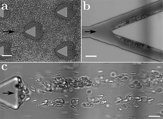
figure : Formation d’agrégats dans une puce microfluidique mimant la microcirculation. (a) Formation d’agrégats de globules rouges drépanocytaires au niveau de coins prononcés. La flèche indique le sens de l’écoulement et la barre d’échelle représente 50 µm. (b) Agrégats de globules drépanocytaires au niveau d’une bifurcation. La barre d’échelle représente 20 µm. (c) En présence de plasma autologue, les agrégats adoptent une morphologie différente de celle présentée en (a) et forment de longues chaînes qui flottent au milieu du canal. La barre d’échelle représente 20 µm. © Manouk Abkarian


En savoir plus: Microfluidic study of enhanced deposition of sickle cells at acute corners and its possible role in vaso-occlusion. E. Loiseau, G. Massiera, S. Mendez, P. Aguilar Martinez and M. Abkarian. Biophysical Journal, Vol. 108, Issue 11, p2623–2632. dx.doi.org/10.1016/j.bpj.2015.04.018 Contact chercheur Manouk Abkarian Centre de Biochimie Structurale CNRS UMR 5048, Inserm U1054, Université de Montpellier 29 rue de Navacelles 34090 Montpellier Cedex Tel :04 67 41 77 13

Unravelling bacterial motility

During its evolution, the gram-negative bacteria Myxococcus xanthus has developed an original property : the ability to move on surfaces in the absence of extracellular appendages such as flagella or pili. This process, called gliding, is mediated by bacterial Focal adhesion sides (FAs). As in eukaryotic cells, FAs sites temporary create a connection between the surface and the bacterial membrane in order to induce the cell movement. The FA sites contain a molecular machinery containing more than fourteen different proteins, distributed between the various cell compartments : external membrane, periplasm, internal membrane and cytosol.

This work was conducted by a tight collaboration between the group of Marcelo Nöllmann at the CBS and the team of Tâm Mignot at the LCB (CNRS, Marseille). JB Fiche, Laura Faure and León Espinosa are the lead authors.

For this study, several microscopy techniques (2-color TIRF, 3D imaging based on astigmatism, RICM, ...) were used to characterize the architecture and formation of FA sites during cell gliding. We show that the motility complex is divided into three distinct functional groups:

*a molecular motor producing proton motive force for the movement
*an inner membrane/cytosolic group acting as a platform for FAs assembly
*a periplasm/outer membrane group allowing the connection with the surface and the force transmission

We discovered that the motor complex moves intracellularly along a right-handed helical path and, when it becomes stationary at FA sites, it powers a left-handed rotation of the cell around its long axis.

The reason underlying this helical movement of the cell are not fully understood yet. However, we hypothesize that the peptidoglycan (PG) is most probably the best candidate because the glycan strands have a global right-handed helical ordering that could serve as a track to guide the motility complex. Also, the rigidity of the PG layer could act both as a transient anchor for the motor complex and opposing contractions to propel the cell.

Reference:

The mechanism of force transmission at bacterial focal adhesion complexes
Laura M. Faure*, Jean-Bernard Fiche*, Leon Espinosa*, Adrien Ducret, Vivek Anantharaman, Jennifer Luciano, Sébastien Lhospice, Salim T. Islam, Julie Tréguier, Mélanie Sotes, Erkin Kuru, Michael S. Van Nieuwenhze, Yves V. Brun, Olivier Théodoly, Aravind L, Marcelo Nöllmann# & Tâm Mignot#.
[* Co-authors]
[# Co-corresponding authors]
Nature. 2016 Oct 5. doi: 10.1038/nature20121.

DNA segregation super-resolved

To ensure proper growth, bacterial genome must be faithfully segregated so that each daughter cell, resulting from cellular division, inherits an identical copy of the mother cell genetic material. Segregation of bacterial genomes is ensured by an active system, called partition system, allowing the separation and the positioning of newly replicated DNA. By using fluorescence super-resolution microscopy techniques, the team of Marcelo Nollmann, in collaboration with the team of Jean-Yves Bouet at the Laboratoire de microbiologie et génétique moléculaires of Toulouse, reveals a new model in which the nucleoid provides a scaffold to guide the proper segregation of partition complexes. This work is published in the nature magazine Nature Communications.

New publication: "Bacterial partition complexes segregate within the volume of the nucleoid."

Authors: Antoine Le Gall, Diego I. Cattoni, Baptiste Guilhas, Céline Mathieu-Demazière, Laura Oudjedi, Jean-Bernard Fiche, Jérôme Rech, Sara Abrahamsson, Heath Murray, Jean-Yves Bouet & Marcelo Nollmann

Journal: Nature Communications, 2016, July, 7:12107, doi:10.1038/ncomms1210

Host-pathogen recognition by the integrated decoy model

NLR multidomain proteins largely mediate the immune response in plants. These immune receptors are the key elements in the recognition of the pathogen leading to plant susceptibility or resistance. Our collaborators in the BGPI (Biology and Genetics of Plant-Parasite Interactions) of the Inra center in Montpellier are interested in the interactions between rice and the phytopathogenic fungus Magnaporthe oryzae. Rice is the main food source for half of the world's population, and crop sustainability is a major public health issue. For several years, CBS's team "Structure, Dynamics and Function of Biomolecules by NMR" has undertaken a collaboration to better understand the molecular mechanisms involved in host-pathogen recognition. A nuclear magnetic resonance approach as well as in vitro and in vivo tests of protein-protein interactions have been made and allow to propose a model explaining the recognition process that implies in particular the receptor integrated decoy domain. This model suggests that fungus effectors (proteins acting as virulence factors) are recognized by the integrated decoy domain but that this recognition also involves synergy with other domains of the immune receptor.To ensure proper growth, bacterial genome must be faithfully segregated so that each daughter cell, resulting from cellular division, inherits an identical copy of the mother cell genetic material. Segregation of bacterial genomes is ensured by an active system, called partition system, allowing the separation and the positioning of newly replicated DNA. By using fluorescence super-resolution microscopy techniques, the team of Marcelo Nollmann, in collaboration with the team of Jean-Yves Bouet at the Laboratoire de microbiologie et génétique moléculaires of Toulouse, reveals a new model in which the nucleoid provides a scaffold to guide the proper segregation of partition complexes. This work is published in the nature magazine Nature Communications.

Read more:
 

Connexion